Macular Degeneration and Oxidative Stress

Recently I discovered that an online friend had been diagnosed with Age-related Macular Degeneration (AMD). My instant reaction was ‘I have something that I believe will help.’ But I decided to do a little research to be sure I had my facts right.

What is AMD?

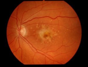
According to the U.S. National Institutes of Health, “AMD is a common eye condition and a leading cause of vision loss among people age 50 and older. It causes damage to the macula, a small spot near the center of the retina and the part of the eye needed for sharp, central vision, which lets us see objects that are straight ahead.

AMD is usually first detected with a standard visual acuity test. Other diagnostic tests (e.g., dilated eye exam, Amsler Grid, Fluorescein Angiogram, Optical Coherence Tomography) may be used to confirm the diagnosis and monitor the progress of the condition.

According to the American Macular Degeneration Foundation, there are two general types of AMD, the “dry” and the “wet” variety.

  • In the “dry” type of macular degeneration, the deterioration of the retina is associated with the formation of small yellow deposits, known as drusen, under the macula. This phenomenon leads to a thinning and drying out of the macula, causing the macula to lose its function. The amount of central vision loss is directly related to the location and amount of retinal thinning caused by the drusen.
  • In the “wet” type of macular degeneration, abnormal blood vessels (known as choroidal neovascularization or CNV) grow under the retina and macula. These new blood vessels may then bleed and leak fluid, causing the macula to bulge or lift up from its normally flat position, thus distorting or destroying central vision. Under these circumstances, vision loss may be rapid and severe.

Apparently there is no known cure for AMD; however there are some treatments that appear to slow its progress, including monthly injections.

Relevance of Oxidative Stress

Research confirms that AMD, as with most chronic diseases, correlates with abnormally low blood glutathione levels – which means the body is experiencing high levels of oxidative stress (Reference 1 and Reference 2). Oxidative stress is a state in which the body has more damaging free radicals than it has the antioxidant capacity to neutralize. High levels of free radicals can do much harm across a variety of the body’s systems, including its visual sensory systems.

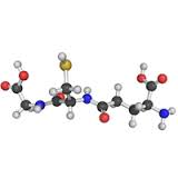
Glutathione (rhymes with ‘glue-da-tie-on’) is a molecule that is produced by every living cell in your body. It is necessary for life in every mammal, including humans. If all of the glutathione were to leave your body, you would not survive two seconds. If you don’t have enough glutathione bad things happen, including disease.

Since glutathione (GSH) is the body’s master antioxidant, a low level of GSH means high oxidative stress. With some exceptions, glutathione is in adequate supply in children. However, from about age 20 onward the natural production of glutathione tends to drop at an average rate of about 10-15 percent per decade. This is a concern because the glutathione molecule is involved in many necessary processes in your body.

In the case of AMD, the “age-related” portion of the name is indicative of the root cause of the condition. The older a person gets the lower their natural intracellular glutathione levels are. AMD is always associated with low glutathione. Therefore it is reasonable to presume that those who are able to keep their intracellular GSH at nominal levels will not develop AMD.

Possible Treatment Option

The natural conclusion this implies is that to slow or stop the progress of the condition in the person who has AMD, they should increase their intracellular glutathione levels substantially. This is a conclusion that was reached in a 2013 study (Reference 3). But as with similar research findings for other chronic diseases, no follow-up work is done to bring this to standard treatment practice. Many folk believe this is because there is no money to be made by the medical and pharmaceutical industries from the use of relatively inexpensive nutritional supplements.

When I heard about my online friend having the “wet” form of AMD, I put a question out to a group of folk that know about glutathione.

Here’s what I asked: Increasing glutathione is supposed to help with macular degeneration. Does anyone know of someone who experienced this effect? I’m unsure what I can tell a friend who I just found out has the condition. Thanks.

After a couple of days, here’s a response I got from a personal friend in the group: Hi John, I have had macular degeneration for many years. My ophthalmologist takes a picture of my left eye every year. It was full of blood spots and lines of blood. Now it is smooth and no blood at all. Big change in about one year. Yeah for <product name>.

The product he named is one that increases intracellular GSH levels. This is one person’s experience. By itself it does not mean this constitutes a confirmed treatment or a cure. But in my mind it is further evidence that the medical community really needs to take a look at increasing intracellular glutathione levels as a bona fide treatment option for chronic diseases like AMD.

References:

Reference 1: Qin L, Mroczkowska SA, Ekart A, Patel SR, Gibson JM, Gherghel D., Patients with early age-related macular degeneration exhibit signs of macro- and micro-vascular disease and abnormal blood glutathione levels, Graefe’s archive for clinical and experimental ophthalmology, (January 2014)

Reference 2: Mrowicka M, Mrowicki J, Szaflik JP, Szaflik MS, Ulińska M, Szaflik J, Majsterek I., Evaluation pro/antioxidant balance in patients with wet form of age-related macular degeneration, Klin Oczna. 2016;118(4):284-8 (original article in Polish)

Reference 3: Zafrilla P, Losada M, Perez A, Caravaca G, Mulero J., Biomarkers of oxidative stress in patients with wet age related macular degeneration, The Journal of Nutrition, Health & Aging. (March 2013)

#amd  #chronicdisease #glutathione

The Impact of ROS on ME/CFS and FMS

This is a topic that is somewhat dear to my heart. My sister was diagnosed with Fibromyalgia a couple of decades ago. And her life has been miserable for much of that time.

Terminology

For clarification, ME refers to Myalgic Encephalomyelitis or Myalgic Encephalopathy, CFS refers to Chronic Fatigue Syndrome, and FMS refers to Fibromyalgia Syndrome (more commonly just Fibromyalgia).

ME/CFS

Chronic Fatigue Syndrome is a term that has been in use for quite some time. A name less recognized in Canada but moving into more common use is myalgic encephalomyelitis (ME). This lengthy name can be easily broken down into its parts, where “myalgic” refers to muscle pain; “encephalo” refers to the brain; “myel” refers to the spinal cord; and “itis” refers to inflammation.

In 2001, Health Canada appointed an international panel of experts in this emerging field of medicine to establish a clinical working-case definition, diagnostic guidelines, and treatment procedures. The panel released a set of guidelines in 2003 and their choice of name to describe the condition was both “myalgic encephalomyelitis” and “chronic fatigue syndrome” – with acronyms shortening it down to a manageable size: ME/CFS.

It is estimated that more than 1 million people in the US population have ME/CFS – 422 per every 100,000 people. It is 4 times as common in women as in men (522 out of 100,000 women have it), and women suffer from ME/CFS in greater numbers than from breast cancer (26 per 100,000), HIV/AIDS (12 per 100,000), or lung cancer (33 per 100,000).

In Canada, Statistics Canada reported in 2005 that over 341,000 Canadians were diagnosed with ME/CFS.

~~ Source: medbroadcast.com

Refer also to a more recent definition from the U.S. Centres for Disease Control and Prevention. Myalgic encephalomyelitis/chronic fatigue syndrome (ME/CFS).

FMS

FMS stands for Fibromyalgia Syndrome. With Fibromyalgia, pain with its devastating and demoralizing effects remains a challenging problem for both patients and care givers. Fibromyalgia is one of the most common diseases affecting the muscles manifested with pain, stiffness, and tenderness of the muscles, tendons, and joints. The painful tissues involved are not accompanied by tissue inflammation. Therefore, despite potentially disabling body pain; patients with fibromyalgia do not develop tissue damage or deformity. The pain of fibromyalgia is generally widespread, involving both sides of the body. Pain usually affects the neck, buttocks, shoulders, arms, the upper back, and the chest. “Tender points” are localized tender areas of the body that can bring on widespread pain and muscle spasm when touched.

FMS typically presents in young or middle-aged females as persistent widespread pain, stiffness, fatigue, disrupted unrefreshing sleep, and cognitive difficulties, often accompanied by multiple other unexplained symptoms, anxiety and/or depression, and functional impairment of daily living activities. There is an overall 6% to 15% prevalence rate in the United States with a five times greater incidence among women than men.

~~ Source: Oman Medical Journal, published May 2012.

ROS

ROS stands for Reactive Oxygen Species, otherwise known as “free radicals.” Oxidative Stress is a condition in which the level of free radicals exceeds the antioxidant capacity of the body – there’s not enough antioxidants to neutralize the radicals and maintain a balance. (Refer to the article Get Rid of the Radicals.) As most people know, radicals are bad for your health. Prolonged periods of oxidative stress lead to disease and disfunction.

Both ME/CFS and FMS exhibit oxidative stress as a common characteristic.

According to a study published in 2014, “Many studies involving peripheral blood measurements have demonstrated significant abnormalities related to increased O&NS [oxidative stress and nitrosative stress] in many patients with ME/CFS.” (Ref. 1)

According to a study published in 2015, “FMS patients have higher oxidative stress index and lower total nitrite levels than healthy controls. In particular, patients with FMS demonstrated higher serum prolidase activity, total oxidative status, and oxidative stress index than healthy controls, and serum prolidase activity positively correlated with pain and fatigue scores.” (Ref. 2)

My Sister’s Story

My sister had been living with a diagnosis of Fibromyalgia for at least a couple of decades. Several days a week she would have difficulty getting out of bed in the morning. Either not enough energy or too much pain or both. And when she did get up and going, everything she did caused pain and drained her energy. This was no way to live. I’m sure I have just glossed over the situation since I have not experienced what she goes through on a daily basis. I could not do justice to her challenges.

A few years ago I became aware of a nutritional supplement that increased the body’s intracellular glutathione levels. And I saw a list of conditions associated with low glutathione. When I saw Fibromyalgia on the list, my attention perked up.

My Mom and my sister live separately but in the same house. And they live about a five hour drive away from where I live. One weekend when I went to visit, I told my sister about the supplement and urged her to try it. I would be her supplier. She agreed – but I think it was because I was her brother and not because she thought it would make a difference.

After I went home I would call my Mom every couple of weeks to see how she was doing. During those calls I would ask about my sister.

“How’s Sue doing?”

My Mom would answer, “Oh, she’s doing alright.”

A few weeks later, “How’s Sue doing?”

“She’s doing alright.”

“How’s Sue doing?”

“She’s doing alright.”

I was starting to get frustrated. This supplement was supposed to help her.

About 8-10 weeks after she started on the supplements, “How’s Sue doing?”

“Oh, she’s had a particularly bad day today.”

I was thinking, “Damn, the stuff was supposed to help.” But before I could say anything, my Mom went on, “You know those pills you gave her?”

“Yeeesss.” I said somewhat hesitantly. I feared she was going to tell me she thought the supplement had caused problems.

Mom said, “Well, her energy levels are up so much and her pain levels are down so much that she’s doing stuff she hasn’t done in 20 years and her muscles are complaining.”

I was shocked. But before I could even think of a response, my mother starts crying and says, “Thank you for giving your sister her life back.”

All I did was suggest that she take some supplements, based on some science research I had done. But I must admit that it felt pretty good.

We later determined that she needed to take more than the standard dose recommended by the manufacturer to see adequate benefits. Things will never get back to the way they were when she was twenty. But they are an order of magnitude better than what they used to be.

Summary

Both ME/CFS and FMS exhibit high levels of oxidative stress (high levels of damaging free radicals). The high level of oxidative stress correlates to high pain and fatigue scores. Glutathione is the body’s master antioxidant. It is produced by the body inside each living cell. Researchers use the level of glutathione as the primary indicator of the level of oxidative stress – the lower the glutathione level the higher the oxidative stress.

Experience shows that increasing intracellular glutathione reduces oxidative stress levels, which leads to a reduction of pain and fatigue. This approach is not a cure; but it may be an effective way to improve the quality of life of those dealing with these conditions.

References:

Reference 1: Gerwyn Morris and Michael Maes, Oxidative and Nitrosative Stress and Immune-Inflammatory Pathways in Patients with Myalgic Encephalomyelitis (ME)/Chronic Fatigue Syndrome (CFS), Current Neuropharmacology, 2014 Mar; 12(2): 168–185. Mar. doi: 10.2174/1570159X11666131120224653

Reference 2: Theoharis C. Theoharides, Irene Tsilioni, Lauren Arbetman, Smaro Panagiotidou, Julia M. Stewart, Rae M. Gleason, and Irwin J. Russell, Fibromyalgia Syndrome in Need of Effective Treatments, The Journal of Pharmacology and Experimental Therapeutics, 2015 Nov; 355(2): 255–263. Published online 2015 Nov. doi: 10.1124/jpet.115.227298

#Fibromyalgia #ChronicFatigue #ME/CFS #glutathione

Thoughts and Observations about PTSD

Post Traumatic Stress Disorder (PTSD) has probably been around for thousands of years. But it is only in the last hundred years or so that it has been recognized as a distinct psychological disorder. Its prevalence first came to public awareness during World War I. Front line soldiers in the trenches of Europe, who were the target of massive artillery barrages, were under constant stress, never knowing whether the next artillery round or bullet had their name on it. And this went on sporadically over several years.

It is not surprising that some men broke under the pressure. If the symptoms manifested at the front line during battle, there was a good chance that the soldier would be charged with ‘cowardice in the face of the enemy’ and summarily shot in front of his comrades as a lesson in discipline. If the thousand yard stare and erratic behaviour occurred after battle, while on leave or after the soldier returned home it was called ‘shell shock.’

The same thing happened in WW II, the Korean Conflict and the Viet Nam War. The condition started to become a matter of public concern in the face of television reporting during and after the war in Viet Nam. This is when some level of research started and ultimately the PTSD designation was applied. It is now recognized as a specific condition by most military forces in the world. In recent years we see more and more veterans of the Gulf Wars and ongoing military operations struggling with PTSD, both in the war zone and after they return home. It has been characterized by an increasing number of suicides among serving members as well as veterans.

But we need to remember that PTSD is not exclusive to the military. The intense traumatic stress that may trigger the condition can occur in civilian populations in a war zone, among people who experience natural disasters, and people who are the victims of sexual abuse or violence. This is particularly true if the stressful situation, over which the person has little control, extends over a protracted period of time.

What Is PTSD?

The U.S. National Institute of Mental Health says this.

It is natural to feel afraid during and after a traumatic situation. Fear triggers many split-second changes in the body to help defend against danger or to avoid it. This “fight-or-flight” response is a typical reaction meant to protect a person from harm. Nearly everyone will experience a range of reactions after trauma, yet most people recover from initial symptoms naturally. Those who continue to experience problems may be diagnosed with PTSD. People who have PTSD may feel stressed or frightened even when they are not in danger.

Symptoms can include:

  • Flashbacks—reliving the trauma over and over, including physical symptoms like a racing heart or sweating
  • Bad dreams
  • Frightening thoughts
  • Staying away from places, events, or objects that are reminders of the traumatic experience
  • Avoiding thoughts or feelings related to the traumatic event
  • Being easily startled
  • Feeling tense or “on edge”
  • Having difficulty sleeping
  • Having angry outbursts
  • Trouble remembering key features of the traumatic event
  • Negative thoughts about oneself or the world
  • Distorted feelings like guilt or blame
  • Loss of interest in enjoyable activities

Other ongoing problems can include:

  • Avoiding relationships
  • Substance abuse
  • Panic disorder
  • Depression
  • Feeling suicidal

It’s More Than a Psychological Problem

The root cause of PTSD is a specific or ongoing traumatic stress-inducing event. We experience some level of stress every day. Some situations are more stressful than others. But traumatic stress raises the bar.

Cortisol is called the stress hormone because it is produced in response to a perceived threat. Cortisol’s effects are particularly noted in the brain, which make sense since the brain is the body’s control centre for the fight-or-flight response to the perceived threat. Many studies over the years have confirmed that prolonged increases in cortisol levels result in increased levels of free radicals. Therefore there is a risk that these radicals will cause damage to brain cells and, if significant enough, may result in altered brain function.

A recent study, involving military personnel as the subjects of the study, concluded that “oxidative stress, increased free radical level beyond excitotoxity, is a possible causal factor for clinical manifestation of PTSD.” (Ref. 1) Since many people with PTSD experience ongoing stress as a result of their symptoms, including re-experiencing the original stressful event, we can presume that additional damage may continue to be done if the level of free radical production due to the cortisol generation exceeds the antioxidant capacity of the subject. (Note: Oxidative Stress is a condition in which the level of damaging free radicals exceeds the anti-oxidant capacity of the body to neutralize them. A state of oxidative stress causes damage to the body’s cells and, if it persists, disease results.)

A Possible Way Forward

Current treatment involves medications and psychotherapy. People with PTSD are advised to seek competent professional help. Having a support network available and taking advantage of it is also strongly recommended.

However, given the possibility that PTSD symptoms are to some degree a result of physical brain chemistry issues due to oxidative stress, one wonders if tackling the oxidative stress situation head on might produce positive results. Increasing the body’s level of intracellular glutathione may be the key. Glutathione is the body’s master anti-oxidant and it is produced in every living cell in the body. Having optimum glutathione levels reduces or eliminates oxidative stress. There are several different ways to help the body increase its glutathione levels. Some are more effective than others; but none involve the administration of pharmaceutical drugs.

References:

Reference 1: Vladimirs Voicehovskis, et al, Oxidative Stress Parameters In Post-traumatic Stress Disorder Risk Group Patients, Proceedings of The Latvian Academy of Sciences, published 2012, Section B, Vol. 66 (2012), No. 6 (681), pp. 242–250. DOI: 10.2478/v10046-012-0016-x

#ptsd #healthscience #glutathione

A Good Night’s Sleep

woman-sleeping-with-smile 1000x560If you suffer from insomnia or if you do not awaken refreshed each morning ready to face the day, you need to pay attention to this.

According to the National Institutes of Health, sleep deficiency is a broad concept that occurs (a) if an individual does not get enough sleep (sleep deprivation), (b) if an individual’s sleeping habits are out of sync with the body’s natural circadian rhythm (sleeping during the wrong time of the day), and (c) if the quality or quantity of sleep is diminished due to a sleep disorder or external factors.

In a 2013 study, the National Sleep Foundation reported that less than half of all Americans (44%) and Canadians (49%) receive a good night’s sleep almost every night. This is a significant health issue.

Adequate Sleep is Important

Sleep is essential to human health. According to a study published in 2005 (Ref. 1) sleep deprivation results in the potential for cell injury and disease due primarily to the resulting reduction in antioxidant levels, particularly glutathione.

Catalase activity and glutathione content, which normally are tightly regulated, were both decreased in liver by 23–36% by 5 and 10 days of sleep deprivation. Such levels are associated with impaired health in other animal models of oxidative stress-associated disease. The decreases were accompanied by markers of generalized cell injury and absence of responses by the other enzymatic antioxidants under study. Enzymatic activities in the heart indicated an increased rate of oxidative pentose phosphate pathway activity during sleep deprivation.

Lack of sleep could increase risk factors for heart disease. According to a 2015 study (Ref. 2):

Decreased quantity and quality of sleep, whether due to sleep disorders or just through lack of proper sleep patterns, have been linked to cardiovascular disease (CVD) risk factors, such as hypertension, obesity, diabetes, and dyslipidemia. Studies have shown that short durations of sleep are associated with greater risk of developing or dying from CVD.

Furthermore, REM (Rapid Eye Movement) sleep is essential to memory and cognitive function. According to a study published in 2009 (Ref. 3):

Compared with quiet rest and non-REM sleep, REM enhanced the formation of associative networks and the integration of unassociated information. Furthermore, these REM sleep benefits were not the result of an improved memory for the primed items. This study shows that compared with quiet rest and non-REM sleep, REM enhances the integration of unassociated information for creative problem solving.

As a side note, intentional sleep deprivation is used as an interrogation technique by police and military forces around the world to take advantage of this phenomenon. There is evidence to suggest that sleep deprivation can lead to false confessions.

REM sleep, however, only occurs during longer periods of deep sleep. It is also during REM sleep cycles that many of the body’s self repair functions are undertaken. People who experience REM sleep tend to dream; but perhaps more importantly, they tend to wake up refreshed and energetic.

What Can You Do?

I would offer four concrete suggestions to improve your sleep situation: avoid stimulants late in the day, schedule your sleep, increase your glutathione levels and get rid of blue light at bed time. Let’s take a look at each of these.

Avoid Stimulants

This is rather obvious but needs to be stated. Many people are in the habit of having coffee or tea after their evening meal. Nicotine (e.g., from smoking) is also a stimulant. But if there is a stimulant like caffeine floating around in their blood stream when it’s time to go to bed, people are unlikely to feel the need to go to bed; when they do go to bed, they are unlikely to go to sleep quickly; and their sleep may not be as restful as it needs to be.

Recommendation: Avoid consuming stimulants in any form for at least eight hours before bed time.

Schedule Sleep

I like to read and do so often in the evening. I guess I inherited that from my dad. He would read Westerns by authors like Zane Grey and Louis L’Amour until all hours. When he was reading he was totally absorbed in the book. Mom used to say she could announce the outbreak of World War Three and he would be oblivious.

I’m the same way. But my preferred genre for escaping reality is sappy romances. (Please don’t tell anyone. It would destroy my macho image.) There have been times when reading late at night that pressure on my bladder forced me to become aware of my surroundings again – and I would realize it was two in the morning. I have had to set the alarm on my mobile phone to remind me to go to bed.

REM Sleep Cycles
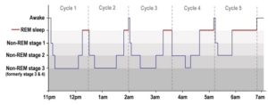
When planning the amount of sleep you need, remember that the beneficial effects of sleep occur during REM sleep cycles. The first cycle lasts about 90 minutes while later cycles last 100-120 minutes. Optimally you should experience five cycles for a good night’s sleep. This forms the scientific basis for the common recommendation that you need 7-8 hours sleep each night.

Recommendation: Determine when you need to get up the next day. Then figure out when you need to get to bed in order to have the time to get an adequate amount of sleep. Then set an alarm for that time so that you actually have a chance of getting your needed sleep.

Remember: An inadequate amount of sleep constitutes sleep deprivation and that is bad for you.

Increase Glutathione Levels

Studies have shown that people with primary insomnia have low levels of glutathione in their bodies. (Ref. 4) If you have low levels of glutathione in your body there is a high probability that, even if you don’t suffer insomnia, you may not experience restful sleep. People with normal levels of glutathione tend to experience REM sleep cycles and awaken refreshed. From about age 20, the body’s natural glutathione production drops by 1 to 1.5% annually. Other environmental factors further diminish the level of available glutathione in most people. Therefore it is not surprising that so many report inadequate sleep patterns.

Recommendation: One important way to counter this situation is to increase your body’s intracellular glutathione levels.

Avoid Blue Light

As I mentioned, I like to read late in the evening to relax. I used to read physical books. But in the past few years I have taken to reading e-books on my laptop or on my phone instead. Coincidentally I have quite frequently found myself reading well into the early hours of the morning without feeling sleepy. This was disastrous when I had morning business meetings. Having my morning alarm sound after only a couple of hours sleep was not pleasant.

I have had reason to monitor my blood pressure more diligently over the last few months (following a cardiac event). My blood pressure tended to stay in a good range. But I noticed a blood pressure increase later in the day on many days. I worked with some professionals to try to get a handle on what was going on but the results of our analysis have so far been inconclusive. During the course of this, one of the health professionals that was helping with this analysis mentioned that there is a correlation of sleep deprivation to higher blood pressure.

Serendipity. I came across an article that shed some light on the issue (no pun intended). The following is an extract from an article entitled Sleep Disorders by Lisa Marshall in the July/August 2016 issue of Discover Magazine.

As recently as the 1980s, researchers assumed the human sleep-wake cycle was not sensitive to light, recalls Charles Czeisler, chief of the sleep and circadian disorders division at Brigham and Women’s Hospital. “In reality it is the most important synchronizer of human circadian rhythms.” In the 80s, Czeisler discovered that specialized ganglion cells in the retina are finely tuned to tell the brain to cut melatonin production when they are hit by a short wavelength, (around 480 nanometers) – precisely that of morning light. Unfortunately, most phone and tablet screens and LEDs emit a similar bluish wavelength, making them exponentially more potent than older yellowish-orange incandescent bulbs. One 2014 study found sleep lab subjects who read from an iPad before bed say nighttime melatonin levels plummet 55 percent after five days (paper book readers saw no reduction). They also took longer to fall asleep, had less REM-stage sleep and were groggy in the morning.

sleeping-man-iphone-apple-bed 1300x867This mirrored my experience pretty closely. So what could I do?

I discovered that my laptop and my phone had a setting that would remove this blue spectrum from the display for selected time periods. I turned this setting on and then proceeded to follow my normal reading patterns. What I discovered was that I would get sleepy while reading on my laptop well before midnight (a change from the past several years). And when I went to bed I was able to get to sleep much more quickly and my sleep was more rejuvenating.

Recommendation: Go to the display settings for your electronic devices and set the Night Shift setting to go on at sunset and off at sunrise.

Summary

I’m starting to sleep pretty well.

  • I haven’t had coffee on a regular basis for several years and I don’t smoke. So I have managed to avoid stimulants before bed.
  • I have my alarm set on my phone to remind me when it’s time to go to bed.
  • I take a nutritional supplement that allows my body to produce glutathione on demand so my glutathione levels are much higher than would be expected for someone my age.
  • And finally I avoid exposure to the troublesome blue spectrum light from my electronic devices in the hours leading to bed time.

Let me know what your experience has been.

References:

Reference 1: Carol A. Everson, Christa D. Laatsch, and Neil Hogg, Antioxidant defense responses to sleep loss and sleep recovery, American Journal of Physiology – Regulatory, Integrative and Comparative Physiology, Published 1 February 2005 Vol. 288 no. 2, R374-R383 DOI: 10.1152/ajpregu.00565.2004

Reference 2: Michelle Kohansieh and Amgad N. Makaryus, Sleep Deficiency and Deprivation Leading to Cardiovascular Disease, Published May 2015, International Journal of Hypertension, Volume 2015 (2015), Article ID 615681

Reference 3: Denise J. Cai, Sarnoff A. Mednick, Elizabeth M. Harrison, Jennifer C. Kanady and Sara C. Mednick, REM, not incubation, improves creativity by priming associative networks, Proceedings of the National Academy of Sciences of the United States of America, Published 23 June 2009, vol. 106 no. 25 10130-10134, 10.1073/pnas.0900271106

Reference 4: Mustafa Guleca, Halil Ozkolb, Yavuz Selvic, Yasin Tuluceb, Adem Aydinc, Lutfullah Besirogluc, Pınar Guzel Ozdemirc, Oxidative stress in patients with primary insomnia, Progress in Neuro-Psychopharmacology and Biological Psychiatry, Volume 37, Issue 2, 1 June 2012, Pages 247–251

#sleep #glutathione #health

About Multiple Sclerosis

multiplesclerosisnervedamageschematicAccording to the Mayo Clinic, “Multiple sclerosis (MS) is a potentially disabling disease of the brain and spinal cord (central nervous system). In MS, the immune system attacks the protective sheath (myelin) that covers nerve fibers and causes communication problems between your brain and the rest of your body. Eventually, the disease can cause the nerves themselves to deteriorate or become permanently damaged.”

The MS Society of Canada says that this country has the highest incidence of MS in the world. From Statistics Canada we learn that about 1 in every 350 people has been diagnosed with MS. That is a disturbing number.

Despite decades of research, the cause remains a mystery. Current evidence suggests that lifestyle, environmental, genetic and biological factors may all contribute. MS is unpredictable and can result in symptoms such as extreme fatigue, lack of coordination, weakness, tingling, impaired sensation, vision problems, bladder problems, cognitive impairment and mood changes.

Because there is no known cure, treatments tend to focus on speeding recovery from attacks, slowing the progression of the disease and managing MS symptoms. Different drugs are used to address different specific symptoms. For example, as with asthma the problematic inflammation is treated with corticosteroids. But there is nothing to treat the disease as a whole.

What is known for certain is that all forms of MS are associated with oxidative stress. You can verify this by referencing PubMed. There is a 100% correlation. To confirm this, go to PubMed and search for ‘multiple sclerosis glutathione’ and you will be presented with a list of hundreds of published papers. Each one will identify low levels of glutathione, which correlates to high levels of oxidative stress. Oxidative stress means that the body has more free radicals than it has antioxidants to neutralize them. Free radicals cause harm to the body and produce cellular inflammation.

In a research paper published in February of 2011 entitled “Radical changes in multiple sclerosis pathogenesis” the authors state that “ROS [Reactive Oxygen Species – aka ‘free radicals’] initiate extensive cellular damage and tissue injury. ROS have been implicated in the progression of cancer, cardiovascular disease and neurodegenerative and neuroinflammatory disorders, such as multiple sclerosis (MS).” (Reference 1) The paper goes on to discuss clinical and experimental studies highlighting the therapeutic potential of antioxidant protection in the pathogenesis of MS.

There is some anecdotal evidence claiming that dramatically increasing the body’s intracellular glutathione levels can result in reduction of symptoms. (Glutathione is the body’s master antioxidant.) Instances of healing of some lesions has also been reported. And this may be supported by the fact that glutathione is needed for myelination, the creation of myelin sheaths to protect nerve fibres. But the problem with scattered anecdotal reports, despite the credentials of the neurologist involved, is that these do not carry the weight of formal controlled clinical studies. A few single reports of success do not provide sufficient data to form a generalization in which the medical community can have confidence. Clearly formal studies of this nature need to be initiated.

So if you have Multiple Sclerosis, what can you do in addition to following your doctor’s recommendations? There are a couple of suggestions I would offer.

These suggestions are based on a few facts. If you have MS your body is experiencing oxidative stress. Prolonged periods of oxidative stress result in your health deteriorating. You could develop a variety of chronic ailments from heart disease to glaucoma to cancer. The only open question is the rate at which your health will deteriorate. If you want to change this reality you need to address the oxidative stress.

  • First, pay attention to your diet and lifestyle to look for changes you can make that will reduce the volume of free radicals produced in your body (e.g., quit smoking, eliminate processed meat from your diet, reduce your exposure to environmental toxins).
  • Second, increase the levels of antioxidants in your body to deal with the free radical load. Increasing the antioxidants you ingest is a good start. But by far the most effective way is to increase your body’s production of glutathione in the cells. (Refer to the article Get Rid of the Radicals!)

Finally, push for clinical trials that examine the benefits of antioxidants as a treatment regimen.

#multiplesclerosis #glutathione #antioxidants


Reference 1: Molecular Basis of Multiple Sclerosis, Biochimica et Biophysica Acta (BBA) – Molecular Basis of Disease, Volume 1812, Issue 2, Pages 131-282 (February 2011)